


Case Study
“OpticStudio is very user-friendly, and that saved us a great deal of time in our experiments. There are so many parameters that at first it can be easy to get lost. But after a bit of training, it became very intuitive to use. The online forums and Zemax Knowledgebase articles were very helpful.”
— Aaron Mok, Ph.D. candidate in Biomedical Engineering / Cornell University
Simulation of Scattering Tissue in OpticStudio Validates Ballistic Transmission Measurement of Tissue Samples at the Micron Scale
Multiphoton microscopy (MPM) images and analyzes biological tissue by non-linear excitation of fluorescence molecules in the specimen. With MPM, two or more photons are absorbed simultaneously to excite the fluorescence molecules. In combination with near infrared (nIr) light excitation, it features deep imaging within highly scattered living organisms that allows life scientists to better understand cellular structure and function in biological samples, in-vivo and non-invasively.
Over the past 30 years, MPM, has emerged to enable deep imaging at diffraction-limited resolution within highly scattered biological samples
MPM uses nIr light (>1100 nm) for fluorophore excitation. However, optical transmission of a large number of biological samples at the nIr are often not readily available, and biological samples of interest are usually at a micron scale.
Many commercial spectrophotometers — the optical system that measure light intensity transmitted through the samples from ultraviolet infrared (UV- Ir) — are only compatible to samples at a centimeter scale. Professor Chris Xu, IBM Professor of Engineering in applied and Engineering Physics at Cornell University, and aaron Mok, a graduate student in the research group, needed a solution that could measure optical transmission at nIr into much smaller specimens, including mouse skulls, mosquito and fruit fly cuticles, rat dura, and the skin of Danionella, the fish genus that includes the smallest vertebrates to inhabit fresh water. These samples are of vast interest to neuroscientists that use MPM.
“We wanted the ability to look at very small samples,” said Mok. “Existing spectrophotometers you can buy don’t achieve the micron-level capabilities we needed.
Therefore, we set out to build a new kind of instrument that can allow us to measure the optical properties in those tiny samples with a high degree of accuracy.”
Key Capabilities
- Incorporating AVxcelerate Sensors into its existing tool chain enabled ZF to non-sequential mode for light scattering simulations.
- Definition capabilities for optical transmission.
- Ease of use, including access to online forums and Zemax Knowledgebase.
Results
- Validated the effectiveness of using single-mode fibers to isolate ballistic photons in the tissue spectrometer.
- Generated spatially resolved transmission maps for five types of micron-scale tissue samples.
- Provided a measurement system to the scientific community that helps yield accurate results for tissue analysis at a very small scale.
Measuring Ballistic and Total Transmission for Tissue Sampling in New Ways
To guide the imaging for an MPM microscope, the optical system designed by Mok and Xu needed to observe three key measurements: the ballistic transmission of each sample, which is the amount of light that passes through the sample without scattering and absorption; the total transmission, which is the overall amount of light that goes through the
sample without being absorbed; and the identification of localized micron- scale absorption and scattering hotspots. So, Mok’s project required an examination of the way light scatters in various samples, and creating
a transmission map that can be used to predict localized scattering or absorption hotspots. His goal was to create spatially resolved transmission maps that would reveal transmission heterogeneity from the five sample types over a wide spectral range from 911 nm to 1624 nm, and report total transmission over a spectral range from 450 nm to 1624 nm.
Previous tissue spectrophotometer design used a system of four apertures to measure ballistic transmission, two in front of and two behind the sample, along with an illumination source to measure ballistic transmission, as shown in Figure 1. This setup only measures transmission in ~cm scale without any spatially resolved transmission information.
Mok’s and Xu's proposed setup, on the other hand, used single-mode fiber (SMF) to isolate ballistic photons of transmission measurements. This method had the advantage to efficiently isolate ballistic photons and allow light to focus onto the sample to obtain transmission of a ~25 um spot in sample. However, it required a way of simulating the previous four aperture design to ensure that this new method generated an accurate measurement of the ballistic transmission that was comparable to the previous setup. Figure 2 compares the two designs.
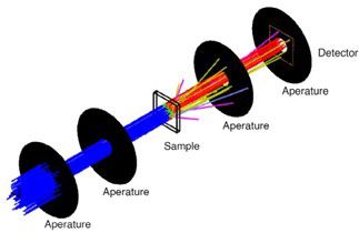
Figure 1. Simulation setup for the double aperture setup in non-sequential mode. Two apertures before the sample ensure that a collimated beam illuminates the sample. The sample is set with an anisotropy factor of 0.9 with different scattering mean path. The distance of the two apertures after the sample are adjusted for an acceptance half-angle of 0.395° or 0.914° to gate out the scattered light. a detector is placed directly after the fourth aperture.
Mok, Xu, and their team performed simulations with ansys Zemax OpticStudio to examine the contribution from non-ballistic photons when using a double-aperture setup at an acceptance half-angle of 0.395° and a published double-aperture setup at an acceptance half-angle of 0.914° (Figure 3).
“We built a simple and versatile system for measuring the ballistic and total transmission of samples customizable for different wavelengths, spatial resolutions, and sample sizes, and the accuracy of the setup is verified with experimental results and OpticStudio simulations,” said Mok. “Using that, we could measure the ballistic and total transmission with a photodetector coupled to a single-mode fiber and an integrating sphere, respectively. It allows a much more versatile setup to measure optical transmission of samples, not just micro-scale biological samples.”
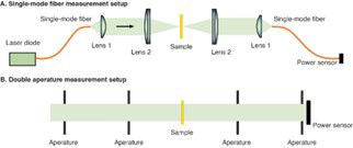
Figure 2. Comparison of the (a) SMF-based micron-scale and (B) double aperture tissue spectrophotometer designs.
Achieving Accurate and Predictable Micron-scale Transmission Results
Successful implementation of the SMF method depended on accurate, reliable simulation. For this reason, OpticStudio played a key role in the team’s success at proving the viability of Mok’s and Xu's design. In particular, OpticStudio’s non-sequential mode enabled them to simulate scattering in biological tissues, and use tools for defining the scattering properties, as well as anisotropic factors for non- isotropic, multi- directional rays.
“Using the measuring method, we created and the simulations in OpticStudio, we verified that the SMF effectively isolates ballistic photons. We measured tissue phantoms of known optical properties, and then comparing the results with theoretical predictions and OpticStudio simulations from a typical double-aperture setup,” Mok said. “We also built a processing pipeline to calculate the transmission map of each sample and extract transmission data from different regions of interest. This led to achieving spatially resolved ballistic and total transmission measurements from microscale biological samples ex vivo, and reported transmission maps to reveal transmission heterogeneity from all of the samples. This measurement method can now be straightforwardly applied to other samples.”
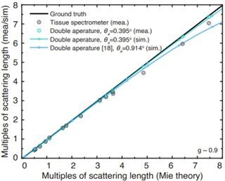
Figure 3. SMF method results showing OpticStudio simulation data (sim) and measurements (mea) and their proximities to the ideal ground truth (solid black line).
Mok also credits other OpticStudio capabilities with helping in key ways. “OpticStudio is very user-friendly, and that saved us a great deal of time in our experiments,” he said. “There are so many parameters that at first it can be easy to get lost. But after a bit of training, OpticStudio became very intuitive to use. The online forums and Zemax Knowledgebase articles were very helpful.”
The ability to document their findings and to back up the accuracy and impact of the simulations in OpticStudio enabled Mok, Xu, and their team to make a convincing case within the scientific and commercial optics community for their new method of isolating ballistic photons. By contributing to these improvements in the micron-scale quantitative imaging of biological phenomena, the team’s innovations and findings will undoubtedly lead to new ways of better exploring the natural world.
Scopri cosa Ansys può fare per te
Scopri cosa Ansys può fare per te
Contact Us
Contattaci subito